Imaging can support the diagnosis of osteoarthritis, show how much a joint has changed, and rule out other problems. Osteoarthritis is mainly diagnosed based on your symptoms and physical exam, with X-rays often providing sufficient imaging evidence when the clinical picture fits. In this guide, we explain how X-rays, MRI, ultrasound, and CT scans may be used, what each test can and can’t show, and how OrthoNJ uses imaging along with your symptoms and exam to guide care.
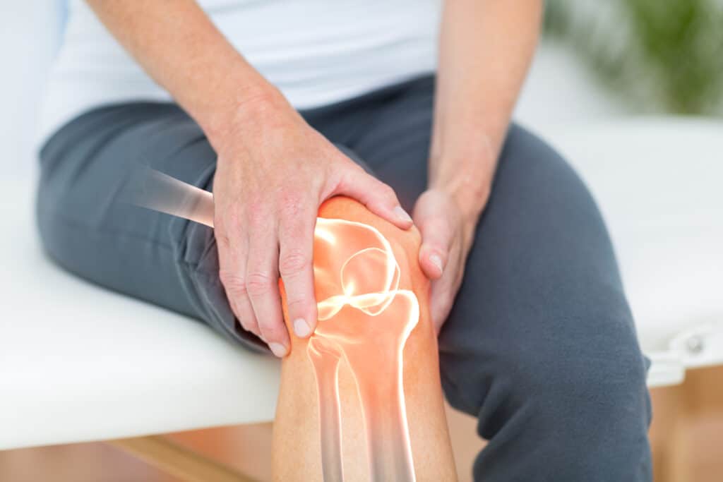
Osteoarthritis is a form of joint wear that affects cartilage, bone, and other structures in and around the joint. Imaging gives your doctor a clearer picture of these changes, but it’s only one part of the evaluation.
Your symptoms, medical history, and physical exam still matter most. Some people have significant changes on imaging and little pain, while others have bothersome symptoms even when imaging findings seem mild.
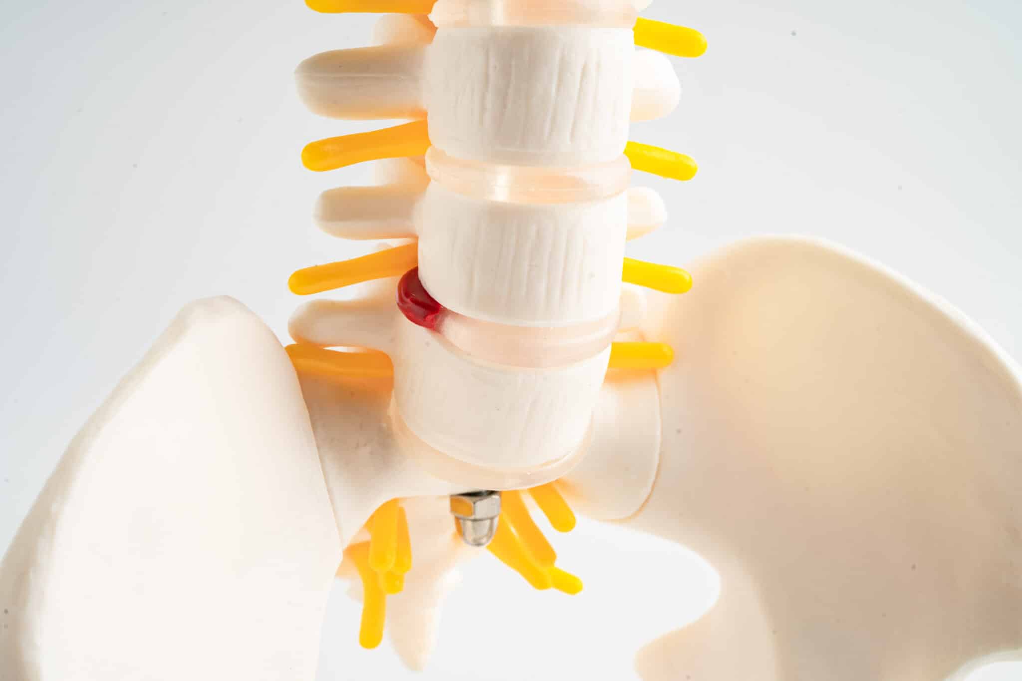
When osteoarthritis is present, imaging may show several typical joint changes. These findings help support the diagnosis and can also help track progression over time.
These findings may appear in different patterns depending on the joint involved, such as the knee, hip, hand, shoulder, or spine.
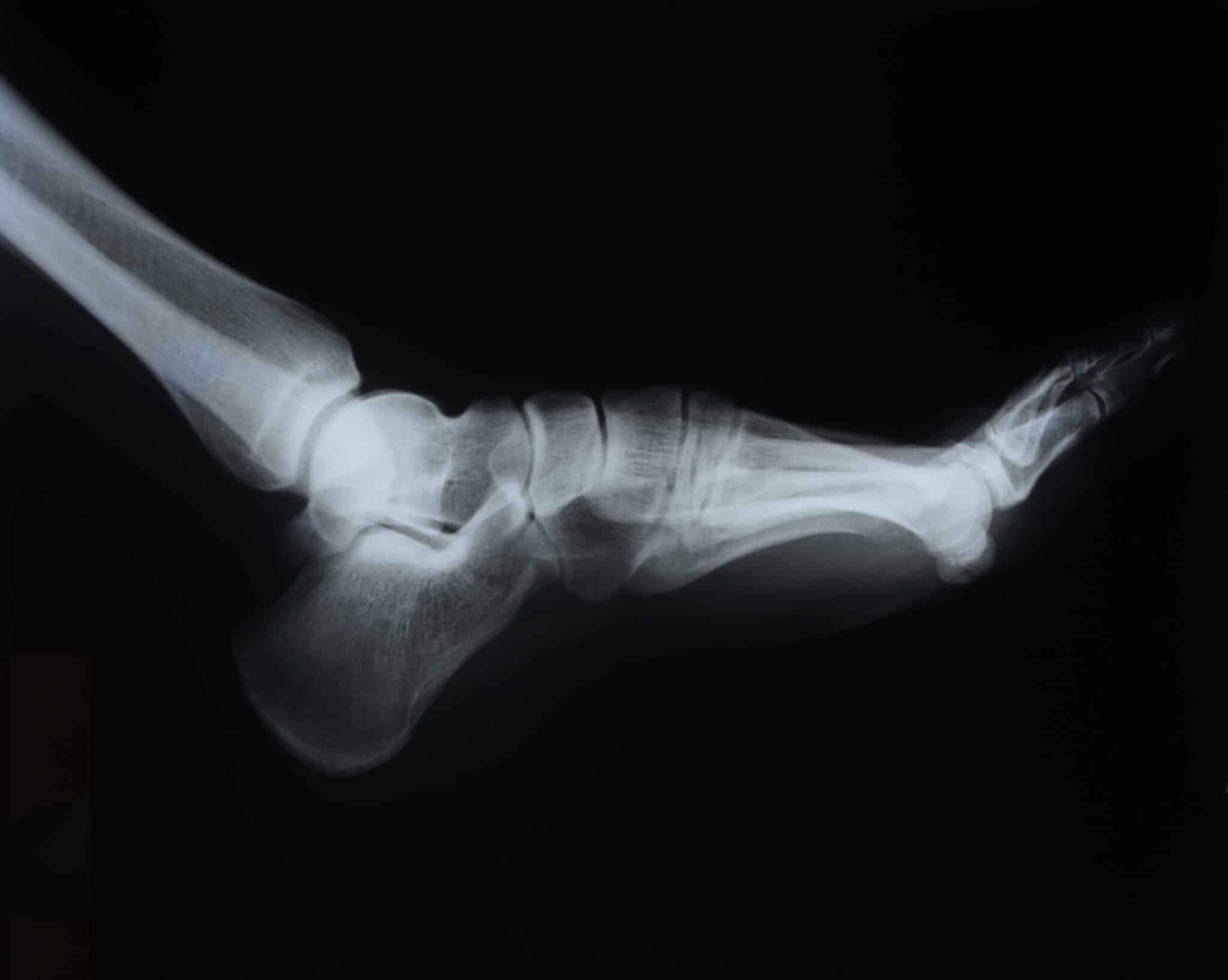
For most patients, plain X-rays are the starting point when osteoarthritis is suspected. They are widely available, quick, and very useful for showing the bony changes that commonly occur with arthritis.
X-rays don’t show cartilage directly, but they can reveal joint space narrowing and other changes linked to cartilage wear. Weight-bearing X-rays, where you stand during the image, can be especially helpful for joints like the knee because they show how the joint behaves under normal load.
X-rays are often enough to support the diagnosis of osteoarthritis when the history and exam fit the picture. They can also help your doctor estimate how advanced the arthritis is and whether certain parts of the joint are more affected than others.
| What X-rays do well | What X-rays don’t show well |
|---|---|
| Show bone spurs and joint space narrowing | Cartilage directly |
| Reveal changes in alignment and joint shape | Ligaments and tendons |
| Help assess severity in many joints | Early soft tissue inflammation |
| Can identify other bone problems | Small details inside soft tissues |
MRI is usually not needed for typical osteoarthritis when X-rays and the physical exam already match the symptoms. Still, it can be useful in selected situations.
An MRI gives a more detailed view of soft tissues and can show cartilage, menisci, ligaments, tendons, bone marrow, and joint lining. This is helpful when symptoms do not match the X-ray findings or when your doctor wants to look for another cause of pain.
MRI often detects more than one issue in a joint, including age-related changes that may not be causing symptoms. That can be helpful, but it also means findings must be interpreted carefully. Not every abnormality seen on MRI is the source of your pain.
Ultrasound is less commonly used as the main imaging test for osteoarthritis, but it can still play an important role. It allows doctors to see certain soft tissue structures in real time and can show joint fluid, inflammation, or changes near the surface of the joint.
It may be especially useful for joints such as the hand, knee, shoulder, or ankle. Ultrasound can also help guide injections more precisely in some cases. Its usefulness in osteoarthritis evaluation is limited and highly operator-dependent, so it is usually an adjunct rather than a primary diagnostic tool for most OA joints.
CT scans are not usually the first choice for routine osteoarthritis imaging. However, they can be very helpful when a detailed view of bone is needed.
For example, CT may be used to evaluate complex joint anatomy, look closely at bone changes, or assist with preoperative planning. In certain joints, such as the spine or areas with prior injury, CT may add useful detail that doesn’t appear clearly on standard X-rays. Both X-rays and CT scans use ionizing radiation, so doctors order them only when the expected benefit outweighs the exposure.
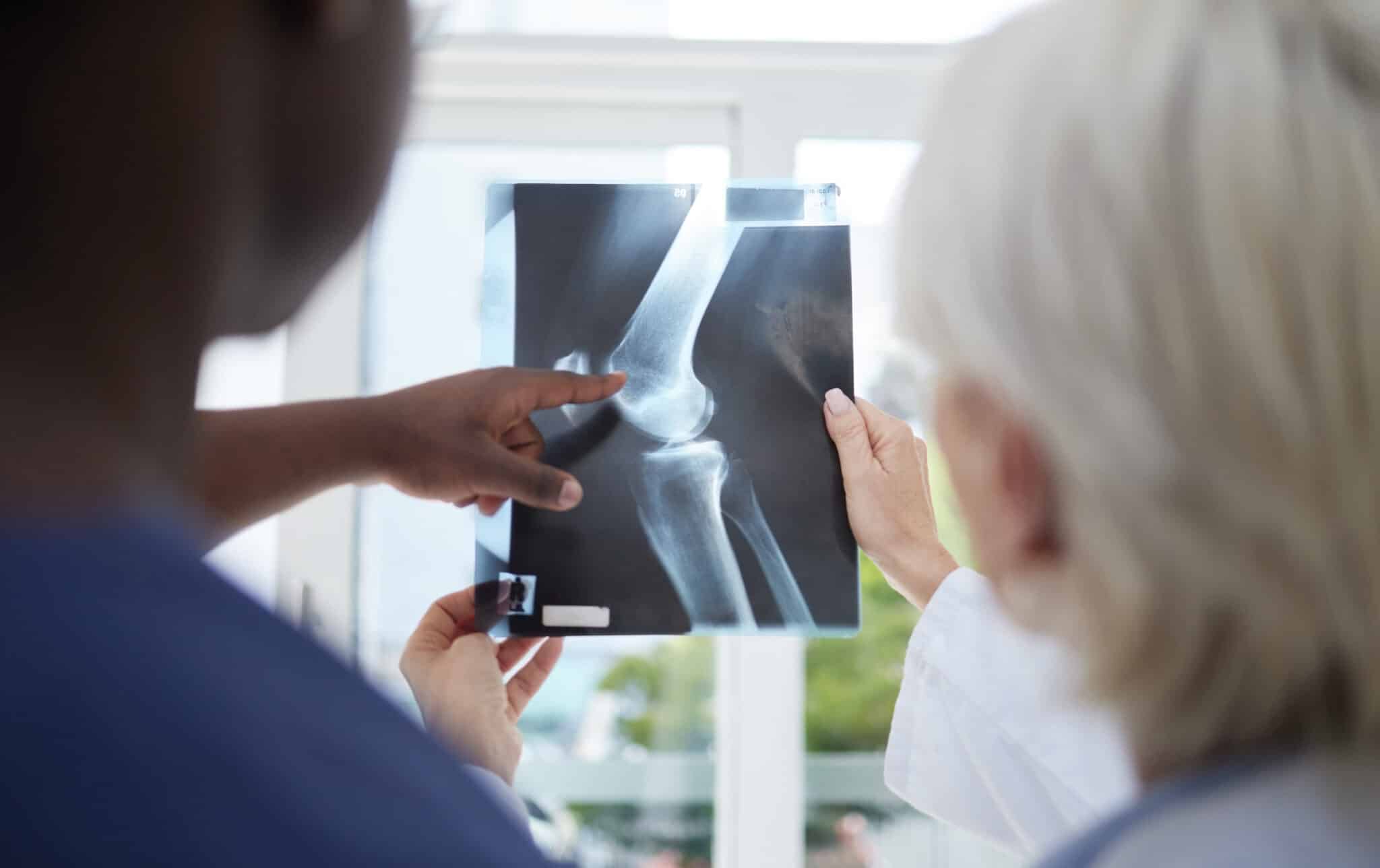
The best imaging test often depends on which joint is causing symptoms. A knee with classic osteoarthritis symptoms may need only weight-bearing X-rays, while a painful hip or shoulder might need additional imaging if the diagnosis is uncertain.
At OrthoNJ, we choose imaging based on your symptoms, exam findings, and treatment goals. That helps avoid unnecessary testing while still making sure important problems aren’t missed.
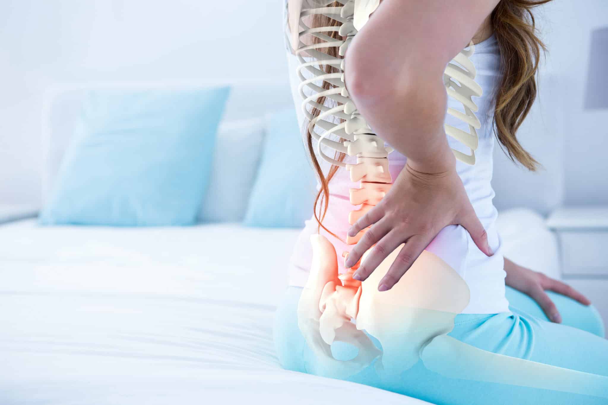
One important point for patients is that imaging and pain don’t always line up perfectly. Some people have major arthritis changes on X-ray and only mild symptoms. Others have a lot of pain with less dramatic imaging findings.
That’s why treatment decisions shouldn’t be based on imaging alone. Your doctor will consider how much pain you have, how the joint functions, what activities you want to return to, and whether other conditions may be contributing.
Imaging can support many parts of osteoarthritis care. It may help support the diagnosis, guide injections, monitor progression, and assist with planning for surgery when needed.
| Imaging test | Best for | Common limitations |
|---|---|---|
| X-ray | Bone spurs, joint space narrowing, alignment, overall arthritis pattern | Doesn’t show cartilage directly or most soft tissues well |
| MRI | Cartilage, ligaments, tendons, menisci, bone marrow, other soft tissue problems | May show findings that aren’t causing symptoms |
| Ultrasound | Joint fluid, surface changes, some soft tissues, injection guidance | Not ideal for seeing deep internal joint structures in every joint |
| CT | Detailed bone anatomy and surgical planning in selected cases | Not usually needed for routine osteoarthritis diagnosis |
You should seek medical care if joint pain is persistent, getting worse, or limiting your daily activities. It’s also important to get checked if the joint becomes very swollen, unstable, or painful after an injury.
If you’re not sure what is causing your joint pain, OrthoNJ can evaluate the problem and decide whether imaging is needed and which type makes the most sense.
Often, X-rays are enough to support the diagnosis when your symptoms and exam fit with osteoarthritis.
An MRI may help if your symptoms suggest another problem, such as a meniscus tear, tendon injury, osteonecrosis, stress fracture, or another condition that isn’t clearly seen on X-ray.
No. Imaging findings and pain levels don’t always match closely. That’s why your doctor looks at the whole picture, not just the scan.
Imaging helps, but it’s only one part of the decision. Surgery depends on your symptoms, function, overall health, and whether other treatments have helped.
This information is for general education and isn’t a substitute for personal medical advice. If you have ongoing joint pain, talk with your OrthoNJ provider about the right evaluation for you.
This treatment info is for informational purposes only. Treatment and recovery vary person to person, and you should consult with your treating physician and team for details on your treatment and recovery process.
Contact one of OrthoNJ's locations spread out through all of New Jersey.