You twist to grab a grocery bag out of the back seat and something low on one side catches. Nothing dramatic. Just a reminder that the small stack of bones between your ribs and your hips carries your whole upper body while letting you bend, lean and turn.
Most people never think about lumbar anatomy until it hurts. Then the questions come fast, usually in the exam room, usually after someone has already looked up a scary word online.
What follows is a plain answer to the questions we hear most: what sits inside your lower back, how discs stay alive without a blood supply, which deep muscles hold the spine steady, why one thin bridge of bone matters to teenage athletes, how the pelvis shares the load, and which symptoms mean you should stop reading and get seen.
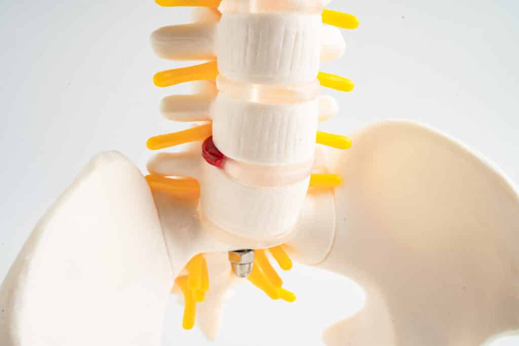
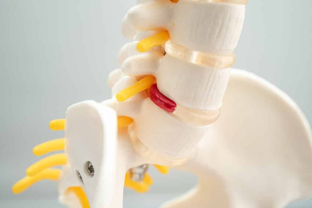
Think of your lumbar spine as a short column with a hollow tube running through the back of it. The column carries weight. The tube protects nerves.
Those lumbar nerve roots carry sensation from the groin all the way to the top of the foot, and they drive the muscles that move your hip, knee and ankle. Two of the lower ones feed into the sciatic nerve. That is why a problem up in your back can be felt down in your calf.
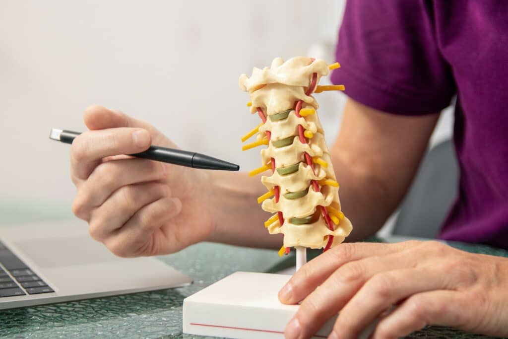
That inward curve is called lordosis, and it is supposed to be there. It balances the weight of everything above it and passes that load down into the pelvis and legs instead of concentrating it in one spot.
Curves can flatten or deepen over time. Posture, old injuries, pregnancy and simple stiffness in the hips can all change how you stand, and a changed curve can shift stress onto the joints and discs. A deep curve by itself is not a diagnosis, though. We look at it alongside how you move and where it hurts.
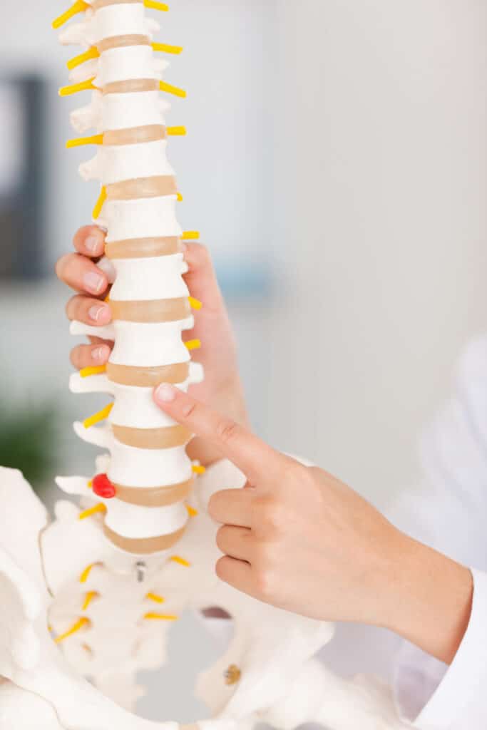
Here is the part most people have never heard. Your discs are the largest structures in the body without their own blood vessels.
Instead, nutrients seep in from tiny vessels in the bone above and below, crossing a thin layer called the endplate, while waste products travel back out the same way. It is slow going. The center of the disc sits the farthest from that supply.
With age, those endplates can harden and calcify, which narrows the path even further. Researchers think reduced transport across the endplate may be one of the things that sets disc degeneration in motion, though it is not the whole story.
Not the ones you see in the mirror. The large back muscles produce movement, but the quiet work of holding each segment in line belongs to a deeper layer.
The multifidus is the one worth knowing by name. It fills the groove on either side of the spinous processes and is best developed in the lumbar region. Close to the bone, it controls single segments rather than swinging the whole trunk. It also attaches near the facet joint capsules, which helps keep that tissue out of harm's way as you move.
In people with long standing back pain, this muscle often looks different on imaging: smaller, with fat showing up where muscle should be. Whether that comes first or follows the pain is still debated. Either way, it is a big reason physical therapy for the lower back spends time on slow control work instead of stretching alone.
It can. Spread across your lower back is a broad, layered sheet of connective tissue called the thoracolumbar fascia. It anchors muscle attachments and passes tension between your trunk, pelvis and legs.
Its layers are separated by looser tissue so they can glide against each other as you bend and twist. In people with chronic low back pain, studies using ultrasound have found less of that gliding motion.
This fascia is also densely supplied with sensory nerve endings, which makes it a genuine candidate for pain rather than a passive wrapper. That matters when imaging of the bones and discs looks unremarkable and you still hurt.
Behind each vertebra is a bony arch. The pars interarticularis is the short bridge of that arch connecting the facet joint above to the facet joint below, and it is the weakest link in the ring.
Repeated arching and twisting can fatigue it into a stress fracture, known as spondylolysis. It shows up most often at the lowest lumbar level, and it is more common in young gymnasts, divers, linemen, dancers and throwers than in the general population. Some people have the defect and never feel a thing, which is why it is sometimes found by accident.
If the crack separates on both sides, the vertebra can shift slightly forward on the one beneath it. That slip is called spondylolisthesis. Most cases in young athletes are managed without surgery, usually with a period away from the aggravating sport followed by a structured rehabilitation program, but this is a topic to bring into the office rather than sort out from a search result.
Worth flagging to us: back pain in a teenager that is worse with arching, lingers past a couple of weeks, or keeps coming back with the same sport.
At the sacroiliac joints. You sit on each side where the triangular sacrum wedges between the two pelvic bones, and thick ligaments lash the whole arrangement together.
These joints barely move. Their job is load transfer and shock absorption, sending force from your upper body into your legs every time you take a step or push up out of a chair.
Pain from this area tends to sit low on one side near the beltline and can spread into the buttock, the groin or the outside of the thigh. It can feel a lot like sciatica, which is exactly why it gets missed. Pregnancy and recent childbirth can make these joints more sensitive, since the ligaments loosen during that time.
That question comes up constantly. The answer is about space.
Nerves in the lower back travel through three separate spaces, and narrowing in any one of them can crowd a nerve:
Narrowing is called stenosis, and it usually builds slowly from a mix of disc bulging, facet joint enlargement and thickening of the yellow ligament. The classic pattern is heaviness, aching or numbness in the buttocks and legs that comes on with walking or standing and eases when you sit or bend forward. Leaning over a cart opens the canal slightly, which is why the produce aisle feels better than the parking lot.
No. It tapers and ends in the upper part of the lumbar region, at a point called the conus medullaris.
Below that, the nerve roots continue downward as a loose bundle named the cauda equina, Latin for horse's tail. They float in fluid inside the canal until each pair reaches its own exit.
This bundle is delicate, and pressure on it is one of the few true emergencies in spine care.
Go to the emergency room if you notice any of the following:
Schedule an exam with your OrthoNJ provider if your pain has not settled after a couple of weeks, if it wakes you at night, if leg symptoms keep returning, or if a young athlete in your house has back pain tied to one sport.
Not the way the phrase suggests. A disc is firmly attached to the bone above and below it, so it does not slide around. What can happen is that the softer inner material pushes through a weak spot in the outer wall.
Sometimes, and sometimes not. Changes in discs, facet joints and the pars can show up on imaging in people with no symptoms at all, so we match what the pictures show against your history and your exam before deciding what is driving the pain.
Gentle movement is usually the better choice for everyday back pain. Long stretches of bed rest tend to leave the deep stabilizing muscles weaker, which can make the next flare harder to recover from.
Because an irritated nerve root sends symptoms along its whole path. A problem at one lumbar level can be felt in the thigh, the calf or the foot, which is one reason we ask you to map exactly where the symptoms travel.
Knowing the parts helps you describe what you feel, and a clear description often shortens the path to the right treatment. Bring us the details: where it starts, what makes it worse, how far it travels, and what you have already tried. Our team will examine your back, talk through what fits, and start with non-surgical care in most cases.
This article is general information about lower back anatomy and is not a substitute for medical advice, diagnosis or treatment. Recovery varies from person to person. For guidance about your own symptoms, schedule an exam with your OrthoNJ provider.
This treatment info is for informational purposes only. Treatment and recovery vary person to person, and you should consult with your treating physician and team for details on your treatment and recovery process.
Contact one of OrthoNJ's locations spread out through all of New Jersey.